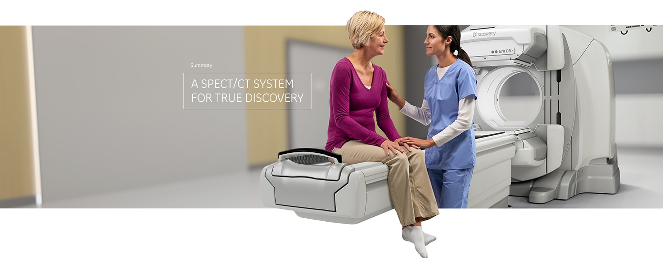
Our mission is to help you in yours. We partnered with people like you to understand what you need from your instruments.
People with the intuition and skills to leverage current technologies and deliver better patient outcomes. Discovery
670 DR is our answer. More than a new imaging product, it’s a system for those dedicated to leading clinical
discovery with hybrid imaging.
1Image quality as defined by an Artifact Index in Phantom testing. Data on file.
2When ASiR is installed, 3D Neuro filter will be disabled.
3In clinical practice, the use of VISR may reduce CT patient dose depending on the clinical task, patient size,
anatomical location and clinical practice. A consultation with a radiologist and a physicist should be made
to determine the appropriate dose to obtain diagnostic image quality for the particular clinical task.
4In clinical practice, the use of ASiR may reduce CT patient dose depending on the clinical task, patient size,
anatomical location and clinical practice. A consultation with a radiologist and a physicist should be made
to determine the appropriate dose to obtain diagnostic image quality for the particular clinical task.
5As compared with same SPECT study corrected by low dose CTAC with standard reconstruction.
6In clinical practice, Evolution options 6a (Evolution for Bone, Evolution for Cardiac, Evolution for Bone Planar)
and Evolution Toolkit 6b are recommended for use following consultation of a Nuclear Medicine physician,
physicist and/or application specialist to determine the appropriate dose or scan time reduction to obtain
diagnostic image quality for a particular clinical task, depending on the protocol adopted by the clinical
site.
6aEvolution Options - Evolution claims are supported by simulation of count statistics using default factory
protocols and imaging of 99mTc based radiotracers with LEHR collimator on anthropomorphic phantom or realistic
NCAT – SIMSET phantom followed by quantitative and qualitative images comparison.
6bEvolution Toolkit - Evolution Toolkit claims are supported by simulation of full count statistics using lesion
simulation phantom images based on various radiotracers and collimators and by showing that SPECT image quality
reconstructed with Evolution Toolkit provide equivalent clinical information but have better signal-to-noise,
contrast, and lesion resolution compared to the images reconstructed with FBP / OSEM.
7Quantitative accuracy defined as equivalence* to well counter-measured injected activity in a test phantom.
*Equivalence means <11% difference when comparing measured counts in SPECT studies corrected by Q.AC-reconstructed
CTAC to measured counts in studies corrected by benchmark reconstructed CTAC. Measured counts are defined
as average within identical ROI’s positioned on SPECT reconstructed slices of homogenous 99mTc solution phantom
study.
 Up to
Up to 
 Standalone, MITA Smart Dose compliant

 Platform compatible with
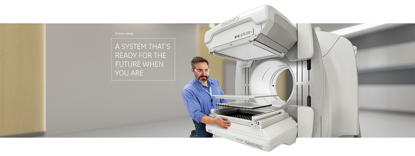
 Pursue
  Digital-detection-ready platform secures your investment with an