Endotracheal Tube Positioning*
As many as 25% of intubations outside of the operating room result in mispositioned ET Tubes, which can lead to severe complications2-6.
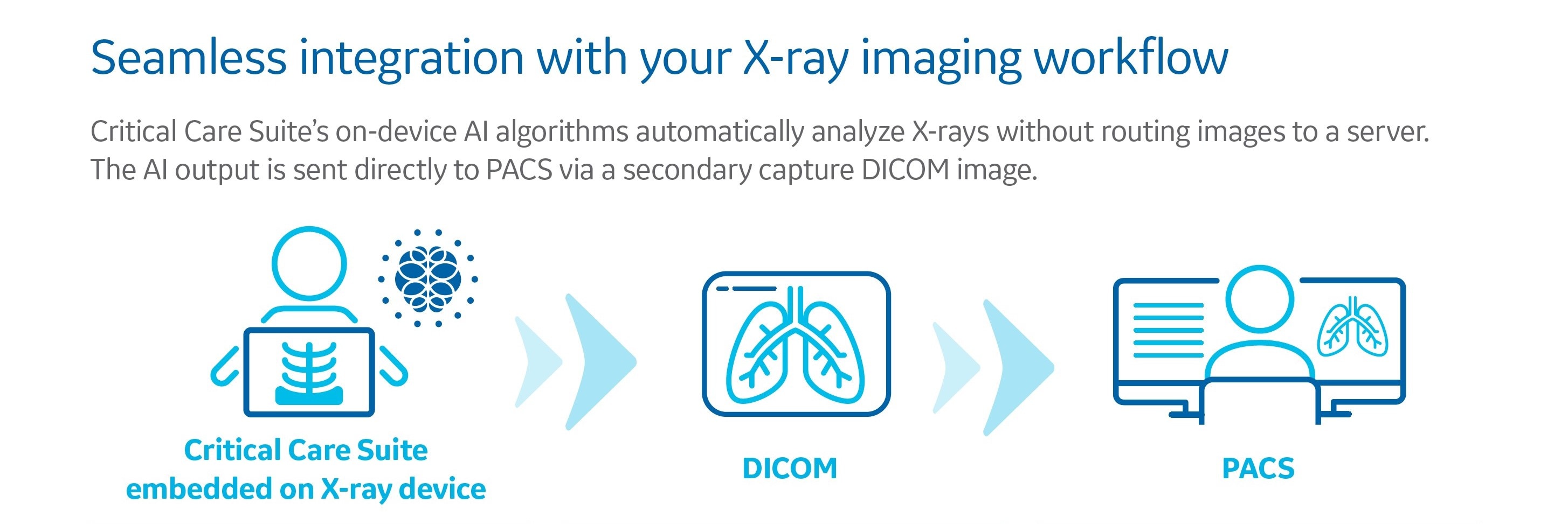
Critical Care Suite’s Endotracheal Tube Positioning AI algorithm is now available on AMX 240 mobile X-ray systems.
Confidence at Point of Care
- Provides an accurate and automated measurement of ET Tube position on the mobile X-ray device within seconds of image acquisition
- The vertical distance of the tube tip and the carina is automatically calculated and displayed on device.
- The average error for the ETT automated measurement is less than 1mm when calculating the vertical distance7.
ET Tube Cases at a Glance
- Enables immediate access to AI-derived measurements in PACS worklist via configurable DICOM® tags
- Displays AI-generated measurements with an image overlay in PACS
Pneumothorax Triage and Notification
On-device AI solution that can help triage critical conditions, such as pneumothorax, alleviating the overwhelming demand that urgent X-rays are placing on radiology teams.
The Pneumothorax Notification is included in Critical Care Suite 2.0 and is available on AMX 240 mobile X-ray systems as well as Definium™ 656 HD and Definium 646 HD fixed X-ray systems.
The Pneumothorax Notification is included in Critical Care Suite 2.0 and is available on AMX 240 mobile X-ray systems as well as Definium™ 656 HD and Definium 646 HD fixed X-ray systems.
High Accuracy8
- Detects nearly all large pneumothoraces (96% sensitivity).
- Identifies 3 out of 4 small pneumothoraces (75% sensitivity).
- Limits false alerts (94% specificity).
- An Area Under Curve (AUC) of 0.96.
Triage Notifications
- Sends a secondary capture DICOM image to PACS and presents the AI results to the radiologist
- Image flags help enable worklist prioritization and have the potential to expedite review of critical findings

Prof Fergus GleesonCMO NCIMI, Consultant Radiologist, Head of Academic Radiology, Director of the Oxford Imaging Trials Unit, Divisional Director for the Oxford University Hospitals NHS Trust, UK

Dr Alex NovakConsultant in Emergency Medicine and Ambulatory CareOxford University Hospitals NHS Trust, UK
Quality Care Suite

Amit Gupta, M.D.Cardiothoracic Radiologist and Modality Director of Diagnostic RadiographyUniversity Hospitals Cleveland Medical Center and Assistant Professor or Radiology at Case Western Reserve University, Cleveland

Dr. Bharat AggarwalDirector of RadiologyMax Super Specialty Hospital, Saket, New Delhi, India

Elizabeth BelcherConsultant Cardiothoracic Surgeon,Oxford University Hospitals, NHS Trust

Steve BarrySenior Radiographer,Oxford University Hospitals NHS Trust, UK
1. Original raw acquired image
2. Intelligent auto rotate
3. Intelligent protocol check
4. Intelligent field of view
5. Critical Care Suite notifications on AMX 240
6. ET tube positioning AI algorithm output
7. Pneumothorax notification
8. Image management with CCS notifications
9. Critical Care Suite secondary DICOM sent to PACS
COVID-19 Case Study
Critical Care Suite enables expedited treatment of a spontaneous pneumothorax in an intubated COVID-19 patient
Supporting Materials
Related products
*510(k) pending. Distributed in accordance with FDA imaging guidance regarding COVID19 public health emergency.
** The technologist notification is generated 15 mins after exam closure. It is contextual and does not provide any diagnostic information. The on-device, technologist notification is not intended to inform any clinical decision, prioritization, or action.
** The technologist notification is generated 15 mins after exam closure. It is contextual and does not provide any diagnostic information. The on-device, technologist notification is not intended to inform any clinical decision, prioritization, or action.
- Stephen E. Lapinsky. Endotracheal intubation in the ICU. Crit Care. 2015; 19(1): 258.
- Jemmett ME, Kendal KM, Fourre MW, Burton JH. Unrecognized misplacement of endotracheal tubes in a mixed urban to rural emergency medical services setting. Acad Emerg Med 2003;10:961–5.
- Katz SH, Falk JL. Misplaced endotracheal tubes by paramedics in an urban emergency medical services system. Ann Emerg Med 2001;37:32–7.
- Lotano R, Gerber D, Aseron C, Santarelli R, Pratter M. Utility of postintubation chest radiographs in the intensive care unit. Crit Care 2000;4:50–3.
- McGillicuddy DC, Babineau MR, Fisher J, Ban K, Sanchez LD. Is a postintubation chest radiograph necessary in the emergency department? Int J Emerg Med 2009;2:247–9.
- Rachh, Pratik, et al. “Reducing STAT Portable Chest Radiograph Turnaround Times: A Pilot Study.” Current problems in diagnostic radiology (2017).
- GE Healthcare Data on File
- FDA 510(k) K183182
WIPRO GE HEALTHCARE PVT LTD
No. 4, Kadugodi Industrial Area,
Bangalore 560067,
Karnataka, India
CIN: U33111KA1990PTC01606
Bangalore 560067,
Karnataka, India
CIN: U33111KA1990PTC01606