


Time is of the essence in the ICU. That's what makes continuous monitoring key.
Patients often present with multiple life-threatening conditions at once. New comorbidities can also emerge quickly, requiring timely medical intervention. Clinicians rely on a variety of imaging and bedside monitoring tools to make quick life-or-death decisions, and their suite of assessment tools increasingly includes critical care ultrasound.
Until recently, ultrasonography was the domain of radiologists, cardiologists, and obstetricians, but the emergence of Point of Care Ultrasound (POCUS) has made the technology accessible to physicians across specialties. Over the past decade, portable ultrasound devices have begun to revolutionize the bedside assessment of ICU patients, giving clinicians real-time clinical insights and a safer way to guide invasive procedures.1 With a growing body of research demonstrating the benefits of critical care ultrasound and endorsements from major medical governing boards, innovative critical care specialists have discovered a variety of novel use cases for POCUS in the ICU.
Use Cases for Critical Care Ultrasound
The more information that critical care clinicians have, the better decisions they can make for patients. To get a look inside those patients, though, clinicians have traditionally needed to rely on radiologists. POCUS lets them obtain crisp, real-time sonographic images on their own at the patient's bedside. In recent years, clinical researchers have described POCUS as the fifth pillar of physical examination that "provides more physiological information than do clinical examination, blood gasses and electrocardiography combined" and predicted that portable ultrasound devices could become as widely used in clinical practice as the stethoscope.2
Modern clinicians rely on POCUS for real-time guidance during potentially risky procedures such as intubation and central line placement as well as to evaluate and manage medically complex patients. In ICUs that routinely leverage POCUS, hemodynamically unstable patients often receive whole-body ultrasound of the heart, lungs, abdomen, kidneys, and vascular system to help determine the sources of their illness.3 With one bedside diagnostic tool, clinicians can quickly assess patients for a plethora of life-threatening entities and gain clinical insights that help guide their care plans.
Lung Ultrasound
Lung ultrasound is a rapid, radiation free modality and when performed with a focused assessment, allows the clinician to rule in or out quickly and accurately diagnose certain clinical conditions.
Burton L, Bhargava V, and Kong M.
Frontiers in Pediatrics
Lung ultrasonography lets clinicians quickly visualize pulmonary injuries and abnormalities. Using lung POCUS, clinicians can detect signs of acute respiratory failure, which may be caused by pneumonia, bronchitis, and asthma exacerbation.1 These conditions have traditionally been diagnosed using chest X-ray (CXR), but numerous studies suggest POCUS could provide initial diagnostic insights very quickly at the point of examination, especially in children. In one recent study published in Frontiers in Pediatrics, researchers explain, "However, the CXR has been found to have relatively low sensitivity and specificity in differentiating etiologies in pediatric acute respiratory failure, suggesting the need for a tool with better diagnostic values. Lung ultrasound is a rapid, radiation free modality and when performed with a focused assessment, allows the clinician to rule in or out quickly and accurately diagnose certain clinical conditions."1 In both adult and pediatric patients, POCUS can also help rule out pneumothorax and assess patients for pulmonary edema, interstitial syndrome, pleural effusion, and lung consolidation. 3
Cardiac and Vascular Ultrasound
Cardiac ultrasound provides insight into a patient's cardiac output, fluid status, and valvular pathology, which can support clinicians as they determine whether heart conditions are contributing to a patient's instability and whether the patient's cardiac function requires further evaluation or special considerations during care.4 It can help indicate hypovolemic shock, pericardial effusion, pulmonary embolism, cardiac tamponade, and other serious conditions that require immediate attention. Vascular ultrasound of the lower extremities can also assist care teams in detecting deep vein thrombosis (DVT), which occurs in approximately 12% of ICU patients and may indicate a pulmonary embolism.3
Renal Ultrasound
Renal failure is common in critically ill patients, affecting roughly 15% to 35% of people admitted into the ICU.3 Using POCUS, clinicians can assess patients for hydronephrosis caused by obstructive uropathy, bladder distension caused by outlet obstruction, and in some cases even identify infected ureteral stones in need of removal. Clinicians can also use POCUS to assess the bladder for urine volume before placing foley catheters, thus reducing the risk of causing urinary tract infection.
Abdominal Ultrasound
Using POCUS, clinicians can detect free fluid in the abdomens of trauma patients and gain other clinically relevant information about a patient's pathology. Abdominal ultrasonography can also be used to screen patients for aneurysm and aortic dissection.
Procedural Ultrasound
POCUS provides clinicians with real-time guidance during bedside procedures.1 For example, it can help them find the ideal location for lumbar punctures and intubate patients with difficult airways. The technology can further guide the placement of central venous catheters and vascular cannulation central lines as well as aid in the drainage of thoracentesis, paracentesis, chest tubes, and abdominal drains.
Best Practices for Use Cases
Critical care practitioners and researchers have spent the past decade exploring the diverse utilities of POCUS. In June 2016, the Society of Critical Care Medicine (SCCM) introduced bedside ultrasonography guidelines for the subspeciality.5 Employing evidence-based insights to guide their recommendations, the SCCM endorses POCUS in a variety of use cases, including diagnosing pneumothorax, DVT, acalculous cholecystitis, renal failure, and interstitial and parenchymal lung diseases. The SCCM also recommends critical care ultrasound for various cardiac and vascular insights.5 However, for more advanced use cases, radiologist assistance may be required.4
Benefits of Critical Care Ultrasound
POCUS provides a way for ICU clinicians to gather information in a timely manner to intervene during life-threatening situations. An ultrasound-trained intensivist can perform a whole-body ultrasound in less than eight minutes and gain an accurate image of the pathology of the patient's major organ systems.3 According to the SSCM, POCUS can identify pleural effusion and DVT with 94% accuracy, making this technique "comparable with, or better than, conventional chest radiography."5 Other recent studies comparing POCUS and CXR have demonstrated the effectiveness ultrasonography as a diagnostic aid. For example, research shows that CXR can identify pleural effusions as low as 175 to 525 ml, but ultrasonography can reveal as little as 5 to 20 ml of fluid with 89% to 100% sensitivity and 96% to 100% specificity. POCUS can also help identify pulmonary edema with 97% sensitivity and 95% specificity.6
According to a growing body of research, POCUS also improves procedural safety. The SSCM finds that the success rate of POCUS-guided paracentesis is 95% vs 61% for image-guided blind paracentesis and reports that most unsuccessful blind taps can be salvaged using ultrasonography. Ultrasound-guided paracentesis has also been shown to decrease the rate of complications and associated costs.1 Meanwhile, the Agency for Health Research and Quality has deemed ultrasound-guided vascular access as one of the 12 most effective ways to prevent medical errors.
Overall, the use of POCUS in ICUs offers opportunities to:
Improve diagnoses.
Reduce the need for supplemental exams.
Decrease the time to initial treatment.
Reduce the likelihood of redundant interventions.2
That adds up to quality care and better patient outcomes.
Challenges of Critical Care Ultrasound
POCUS is becoming an increasingly useful tool for clinicians, but lack of experience can be a real barrier to adoption. Ultrasound image quality and diagnostic reliability depend on the skill of the practitioner, and until recently, clinicians have not generally been trained to use this technology.1 Today, most large residency programs include ultrasound training in their curriculum, and the SCCM offers a certification program to bring untrained physicians up to speed.
Physiological and pathological factors attributable to the patient can also affect image quality and interpretation. ICU patients may be difficult to position and are often connected to ventilators, cardiac monitors, and various bedside telemetry. However, as POCUS devices become easier to maneuver, clinicians can overcome many of these ergonomic challenges.
POCUS equipment can also present an infection risk and potentially spread hospital-acquired infectious diseases. To mitigate these risks, various professional groups have published POCUS disinfection guidelines.
Despite the challenges that remain, critical care ultrasound is rapidly becoming a common standard of practice. While clinicians become increasingly proficient with POCUS, the technology is becoming more advanced and more affordable—opening the door for even more diverse utilities for POCUS in the ICU.
References:
- Burton L, Bhargava V, and Kong M. Point-of-care ultrasound in the pediatric intensive care unit. Frontiers in Pediatrics. 2021; 9:830160. https://www.ncbi.nlm.nih.gov/pmc/articles/PMC8845897/.
- Zieleskiewicz L, Lopez A, Hraiech S, et al. Bedside POCUS during ward emergencies is associated with improved diagnosis and outcome: an observational, prospective, controlled study. Critical Care. 2021; 25:34. https://www.ncbi.nlm.nih.gov/pmc/articles/PMC7825196/.
- Guevarra K and Greenstein Y. Ultrasonography in the critical care unit. Current Cardiology Reports. 2020; 22(11): 145. https://www.ncbi.nlm.nih.gov/pmc/articles/PMC7481757/.
- Levitov A, Frankel H, Blaivas M, et al. Guidelines for the appropriate use of bedside general and cardiac ultrasonography in the evaluation of critically ill patients—part II: cardiac ultrasonography. Critical Care Medicine. June 2016; 44(6).1206-1227. https://journals.lww.com/ccmjournal/Fulltext/2016/06000/Guidelines_for_the_Appropriate_Use_of_Bedside.23.aspx.
- Frankel H, Kirkpatrick A, Elbarbary M, et al. Guidelines for the appropriate use of bedside general and cardiac ultrasonography in the evaluation of critically ill patients—part I: general ultrasonography. Critical Care Medicine. November 2015; 43(11): 2479-2502. https://journals.lww.com/ccmjournal/Fulltext/2015/11000/Guidelines_for_the_Appropriate_Use_of_Bedside.26.aspx
- Taylor I, Anjum F, and O, Rourke, MC. Thoracic and lung ultrasound. StatPearls. https://www.ncbi.nlm.nih.gov/books/NBK500013/. Last updated June 19, 2022.
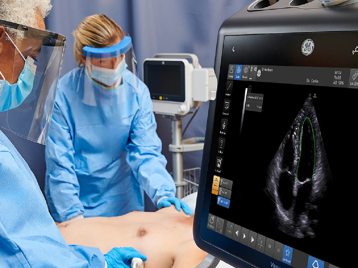
A patient's status can change rapidly in the ICU. Venue products provide critical care ultrasound support for fast care decisions.