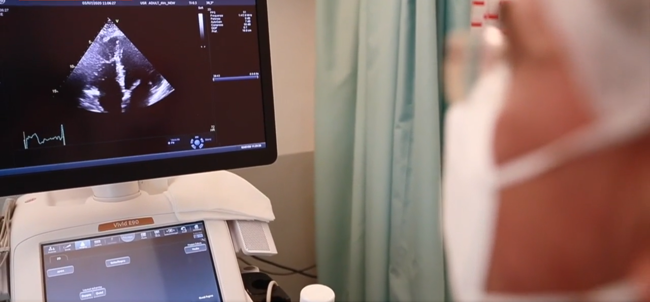
As previously reported[1] in Insights, evidence around the relationship between COVID-19 and the heart are unfolding globally day by day. One developing area points to a connection between COVID-19 and strain on the heart’s right ventricle (RV).
One expert believes that the function of the RV is even more important than the function of the left ventricle (LV) in predicting the prognosis and the functional capacity of those patients who will survive COVID. While the LV is more susceptible to infectious complications of widely understood conditions such as myocarditis, the RV is more of a mystery as evidence shows it’s susceptible to COVID complications due to clots that are forming in the lung, pulmonary embolism or obstruction.
GE Healthcare Insights sat down with Luigi P Badano, MD, PhD, Professor of Cardiology at the University of Milano-Bicocca, Director of the Cardiovascular Imaging Unit at Istituto Auxologico Italiano, IRCCS, and leading expert on RV and strain imaging, to gain a better understanding of his findings.
Q: What have you learned about COVID’s impact on the heart?
Luigi P Badano, MD, PhD: We have learned a lot because we started from knowing nothing and have been learning in the field, so our knowledge improved day by day, week by week. So far, we know that COVID is a disease that mainly involves the inflammatory and the coagulation system. There is a relatively high percentage of patients with involvement of the heart, and most of them have a subclinical involvement. So, the need of new echo techniques able to measure the function of the myocardium—the muscle of the heart—is becoming vital to identify those patients.
Q: Have you seen any linkage between the RV and COVID?
Luigi P Badano, MD, PhD: We started being interested in strain imaging of the free wall of the RV several years ago because, different from the other echo techniques, it provides actual measurements of the function of the RV myocardium. Since many COVID patients have thromboembolic complications, and in a few of them pulmonary embolism, we felt that the RV may be impacted by the COVID complex disease. We started to take measurements to identify those patients who have initial and sometimes subclinical involvement of the RV.
Q: What are some of the complexities in trying to look at the RV?
Luigi P Badano, MD, PhD: The RV is really challenging because we cannot calculate volumes based on linear and area measurements as we do for the left counterpart due to its very peculiar shape. So far, we have just taken surrogate measurements to try to estimate both size and function. The best way to measure the size of the RV is to use three-dimensional echocardiography. When the RV is dilated, we are already in an advanced stage of disease. Strain—the measurements of the function of the myocardium—can detect involvement of the RV well before it becomes dilated and dysfunctional. We know that anticoagulation was one of the most effective treatments for those patients. It is hoped that by being able to detect earlier the cardiac damage, we can improve the prognosis of those patients.
Q: What interesting techniques are you using to identify RV strain with COVID patients?
Luigi P Badano, MD, PhD: Our techniques are mostly linked to the use of 3D and strain echocardiography. Something new that we are doing is to use some computer techniques—sort of radiomics—to see if we can get additional information from analysis of the raw data of the images, something that cannot be seen with the human eye and we are not able to measure with conventional software packages. This is an application of artificial intelligence to the images we get with the ultrasounds. The hope is that those techniques will be able to not only shorten the echo study performed by the sonographer or the doctor, but also to improve our ability to detect subclinical, very initial damages to both the LV or RV.
Q: What happens after you’ve identified strain with 3D ultrasound?
Luigi P Badano, MD, PhD: If the strain of the right ventricle or the pressure of the pulmonary artery are increased, we look at the coagulation system of the patient and eventually we prescribe a CT scan of the lungs to be sure that there is no pulmonary embolism. If not, another potential diagnosis is that myocarditis is affecting one or both ventricles.
Q: What key questions still exist for patients with RV strain and COVID?
Luigi P Badano, MD, PhD: What we need is to have an assessment of the entire wall of the RV because so far what we can explore is really a very small slice of the RV that represents no more than 5-10% of the entire myocardial muscle. If we would be able to assess the entire free wall of the RV—and this would be possible only if we have a 3D strain—then it’s likely to provide much more accuracy and much more sensitivity in our diagnosis. Three-dimensional echocardiography, particularly for the RV, is our preferred technique, but more and more, we are applying the ability of artificial intelligence to provide additional power to our 3D images.
[1] https://www.gehealthcare.com/article/solving-the-link-between-covid-19-and-cardiac-impact


