Clinical Image Gallery
LAVA Flex Axial
3D MERGE Axial
T2 frFSE Sagittal
T1 FLAIR Sagittal
T2 FSE Coronal
T2 PROPELLER Coronal
PD PROPELLER Fat Sat Axial
PD PROPELLER Coronal
3D SWAN Axial
FIESTA Cine Long Axis
3D TOF
Inhance Deltaflow, acquired in 3 stations
Inhance 3D Velocity
VIBRANT Flex Axial
Whole Body imaging with GEM Suite using T1 fsPGR and T2 SSFSE with ARC acceleration.
T2 PROPELLER Sagittal
FIESTA Cine Four chamber view
PD frFSE Fat Sat Coronal
Inhance 3D Inflow IR
PD PROPELLER Fat Sat Sagittal
PD frFSE Axial
PD FSE Fat Sat Coronal
T1 IDEAL Coronal
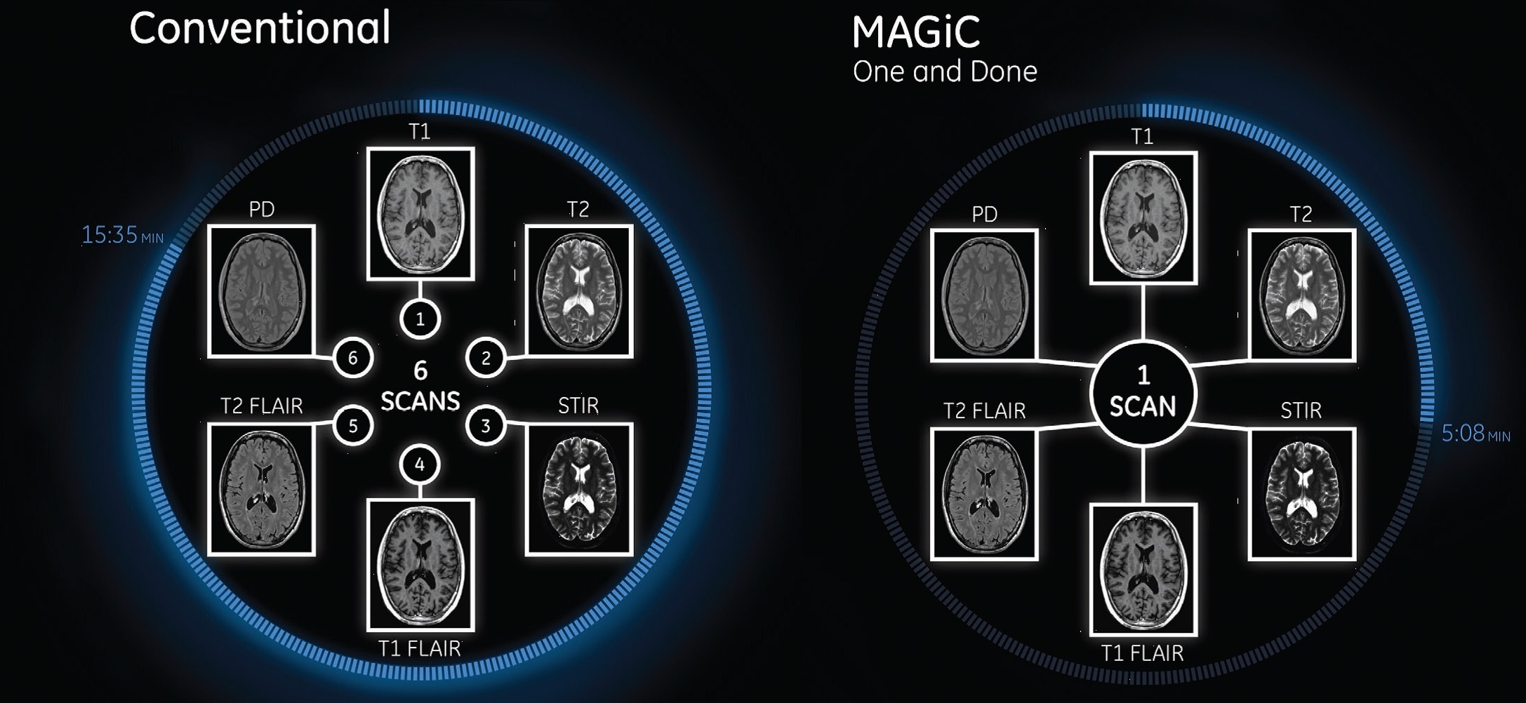
MAGiC (Magnetic Resonance Image Compilation)
Click here to discover the MAGiC Experience
For the first time ever in the industry, you can generate multiple image contrasts in a single MRI scan including T1, T2, STIR, T1 FLAIR, T2 FLAIR* and PD contrasts of the brain in a single acquisition.
One scan that can do the work of many, with images acquired in as little as half the time to acquire separate sequences, fully integrated in a seamless workflow.
*It is recommended to acquire conventional T2 FLAIR images in addition to MAGiC.
MAGiC
Complete flexibility for multiple contrast changes
You can change the contrast of the image by manipulating TR, TE and TI even after the scan is completed and the patient has exited the MR room. MAGiC processing will provide you any desired contrast paving the way to new diagnostic capabilities.
MAGiC even goes beyond standard MR weighted images by processing raw data into parametric T1, T2 and PD maps.
Benefits
See how the Discovery MR750w gives you the experience you deserve, the performance you desire and the versatility you demand.
Its full 3.0T magnet and 70 cm bore work together to generate extraordinary image quality without compromises. The result for clinicians is new levels of diagnostic performance.
Patients benefit from a more comfortable scan experience. Beyond the widened bore and soft, flexible coils, the table surface has been completely re-designed to help alleviate pressure points for a more relaxing exam.
Technologists benefit from an ergonomically-friendly interface that mimics the same consumer-designed devices they use every day, allowing them to focus their attention where it belongs—on their patients.
Technology
Insightful Technology. Thoughtfully Built.
Magnet A new, light-weight magnet with a compact footprint that supports a 70 cm patient gantry and offers a large 50 x 50 x 50 cm useable field of view with excellent homogeneity for uncompromised coverage and quality.
MultiDrive RF Transmit Fully automated and independent RF pulse amplitude and phase control produce consistently clear 3.0T images.
OpTix Optical RF offers high channel count, analog to digital-optical signal conversion where it matters—inside the scan room to minimize noise and signal degradation, but away from the patient.
A large usable field of view is needed to properly image off-center anatomy such as a shoulder or hip. So the Discovery* MR750w features a 70 cm flared, open bore design with a large 50 x 50 x 50 cm field of view.
Gradients and RF body coils are water and air-cooled for optimum duty-cycle performance, short repetition time (TR) and echo time (TE), producing sharp and clear images.
The GEM Suite of flexible coils embrace the patient, bringing both function and comfort while helping to minimize anxiety and motion during the exam.
Intuitive applications help clinicians utilize the full potential of 3.0T MR imaging.
Experience
System Design – Discovery MR750w features an innovative new design inspired by the metaphor of protection.
With technologist workflow and patient experience in mind, key features include:
- 70 cm Patient Bore – Bright inner bore lighting and a flared gantry lead to a comfortable, open experience.
- High Resolution In-Room Operator Control (iROC) – Fast exam set-up with high-resolution color console mounted on the front of the magnet. Easy to see patient, system and scan information, and control and select parameters in real time right in the room.
- Sleek dual-sided controls – Control the scanner from either side of the table. Simplify patient set-up with easy access to cardiac or peripheral gating leads and IV lines.
- IntelliTouch Patient Positioning – Boost exam productivity with IntelliTouch patient positioning, by eliminating the need for laser alignment and reducing the steps to position patients in as little as 30 seconds.
- Sophisticated LED accent lights – The parenthesis accent lights on the sides of the Discovery MR750w not only enhance the overall aesthetic beauty of the system, they express the “caring hands” metaphor of embracing the patient in a comfortable experience.
GEM Suite
The GEM Suite is an integrated system that combines high-density RF surface coils and innovative software technologies designed to provide uncompromised image quality, improved workflow and increased patient comfort to help minimize anxiety and motion.
Key features include feet-first imaging for all anatomies, flexible designs that comfortably embrace the patient, comfort tilt to improve brain and neck exam form, reduced exam times through fewer coil exchanges, and comfortable variable density padding designed to help minimize pressure points. Each component of the GEM Suite can be used individually or combined for complete head-to-toe imaging.
GEM Express Patient Table - Redesigned with an integrated posterior array and variable density pad set, designed to enhance patient comfort, allow for set up outside the exam room, and transport patients in emergency situations.
GEM Posterior Array - Embedded high-density posterior array with optimal coil element geometry that enhances spine, abdomen, cardiac, and lower extremity scanning.
GEM Head and Neck Units - Consisting of four imaging components (a head base-plate, an anterior neuro-vascular face-array, the GEM cervical array, and the open face adaptor), the GEM HNU units may be positioned at either end of the table to support feet-first or head-first imaging. The open-face design provides a patient-friendly, open feel to minimize visual obstructions. For C-spine imaging, or with the open face adapter, the base plate may be used with the innovative GEM cervical array, or with the open face adapter to accommodate large or claustrophobic patients.
Comfort tilt - Elevates the superior end of the GEM head and neck units and is designed to improve comfort while enhancing image quality by positioning the anatomy closer to the coil elements.
GEM Anterior Array - A lightweight, flexible, thin and pre-formed array to embrace patients’ various sizes and shapes. With 54 cm of S/I coverage, the anterior array permits upper abdominal and pelvic imaging without repositioning the patient and supports parallel imaging in all 3 planes.
GEM Peripheral/Vascular Array - A high-density PV / lower extremity array that facilitates imaging of the thighs and lower legs with parallel imaging in all 3 planes. The coil incorporates an innovative self-supporting hinge design between the upper and lower elements to accommodate patients of various sizes with simplified set-up.
Supporting Materials
Related
JB01445IN
Bangalore 560067,
Karnataka, India
CIN: U33111KA1990PTC01606